Conditions Treated
The Conditions Our Team Has Treated
Anal Abscesses/ Anal Fistula
Anorectal abscesses are infections that develop in and around the anus. They usually result in a swelling, constant pain, and sometimes fever. The pain is usually not relieved or made worse by a BM. Anorectal abscesses, if left long enough, will usually spontaneously drain pus through an opening that develops in the overlying skin. The drainage of pus from the infected cavity lessens or relieves the pain and swelling. In some patients, after spontaneous drainage, the abscess fully resolves without further incident. Sometimes, however, the abscess does not fully empty. In this situation the infection persists and continues to drain either on a daily or intermittent basis. Patients with an abscess that has spontaneously drained but persists need to see a surgeon and undergo an incision and drainage.
Ideally, before an abscess has the opportunity to drain spontaneously the patient is examined by a surgeon who, after making the diagnosis, will incise and drain the infection. Therefore, patients who develop constant pain in the area around the anus (with or without swelling or fever) should immediately see a general or colon and rectal surgeon. Before performing the drainage procedure the surgeon will obtain consent after explain the situation as well as the pros, cons, and possible complications of this minor surgery. The skin around the anus and abscess is cleaned and prepared after which a local anesthetic (lidocaine or similar agent) is injected into the skin surrounding the abscess. After the area has been numbed a needle is passed into the suspected abscess to confirm that the swelling contains pus. Next, a scalpel is used to drain the infection through a small incision made over the swelling. The surgeon then probes the area to make sure that all of the pus has been drained after which the cavity is irrigated and then lightly packed with gauze. A dressing is then placed over the area.
Sitz baths should be started after the first BM or about 24 hours after the drainage procedure if there has been no BM (see anorectal postoperative instructions). At the time of the first sitz bath, the packing in the abscess cavity is removed. After the bath a gauze dressing is used to cover the wound. Sitz baths should be carried out 2 to 3 times a day and after BM’s for the first 4 to 7 days. If needed, narcotic containing medications are taken for several days. The alternative is to take Tylenol or advil. It is a good idea to take a stool softener (docusate sodium, 100 mg pills, 3 times/day) during this time period in order to avoid constipation and difficult BM’s.
Antibiotics are not routinely given to abscess patients after surgical drainage. The exceptions to this policy are patients who have diabetes or have suppressed immune systems for other reasons (patients on steroids, transplant patients, etc.); these patients are given antibiotics for 5 to 7 days after abscess drainage.
Unfortunately, in between 33 and 66 % of abscess patients, after either spontaneous or surgical drainage, a fistula forms. Abscess patients, prior to undergoing incision and drainage, need to understand that a fistula may form.
Anal Fistulas
A fistula is a tube or tunnel between two structures. In the case of anal fistulas, the connection runs between the skin surrounding the anus and the anal canal. Anal fistulas have an external (on the skin) and an internal opening (in the anal canal). The cause of the vast majority of anal fistulas are anorectal abscesses. The symptoms associated with anal fistulas include: intermittent swelling and discharge, pain, bleeding, and a lump or small area of hardness next to the anus. The discharge may contain pus or blood and is most often noted between BM's. Because the fistula tract may become clogged or blocked, small abscesses can develop from time to time. These abscesses usually result in a painful swelling. Most often, after a few days, the abscess spontaneously drains through the external oal opening which usually relieves both the swelling and pain. With few exceptions (mainly fistulas due to Crohns Disease or Ulcerative Colitis), once a fistula has formed it remains. After learning that they have an anal fistula most patients, make the decision to undergo surgery with the hopes of curing this problem.
Fistulotomy
The path each fistula takes varies from patient to patient. Some fistulas tunnel beneath the skin alone, however, most run through and across some of the anal sphincter muscle. The amount of muscle lying between the fistula tract and the skin surface is an important factor because the simplest and most effective way to get rid of a fistula is to surgically divide the skin, muscle, and other tissue down to the level of the fistula tract. This procedure, called a fistulotomy, is carried out after a thin wire-like metal probe is inserted through the external opening of the fistula and passed through the tract and out of the internal opening. The fistulotomy "unroofs" the tract and allows all of the infected material in the tract to be curretted and removed. The wound is left open to heal slowly over the course of 2-4 weeks. Of the treatment choices for anal fistulas (see below), fistulotomy has the highest success rate (over 90%).
The downside of fistulotomy is that it requires division of a portion of the sphincter muscle in most patients. When muscle is cut there is a chance that sphincter function will be altered. Thankfully, most patients' sphincter control is not affected by the loss of small or even moderate amounts of muscle, however, a small number of patients will notice after fistulotomy that they have difficulty holding back flatus (gas) or, very rarely, liquid stool. The surgeon must determine, at surgery, how deep the fistula is (meaning how much of the anal sphincter would need to be cut if a fistulotomy was performed). If the fistula involves a large proportion of the sphincter muscle then the fistulotomy operation would not be done. Instead, the surgeon would perform one of the other fistula operations described below that either avoids or minimizes muscle division.
Cutting Seton
This method divides the sphincter muscle within the fistula tract over a period of 2 to 4 weeks instead of immediately in the operating room. It is believed by many surgeons that the chances of developing incontinence is much lower when the muscle is cut slowly over a long period of time. The cutting seton method also gives the patient more control over the treatment because it is possible to remove the seton at any time after surgery if the patient notices any symptoms of sphincter weakness. If the seton is removed early then some of the sphincter muscle in the tract is preserved (as opposed to a fistulotomy in which the muscle in the tract is fully divided at once). A brief description of this method follows.
The first step is to identify the internal and external openings and to pass a probe through the tract. The tract is then cleaned with a curette. Next, the skin lining between the two openings is incised which exposes the underlying sphincter muscle. A thick suture or a thin rubber/plastic string (the seton) is then passed through the fistula tract (after curetting and cleaning) and the two ends are tied together very tightly which places pressure on the muscle. As mentioned, the seton divides the muscle contained within the fistula over a 2 to 4 weeks period. Patients tolerate cutting setons well. In some cases the seton may need to be tightened several weeks after surgery. This can usually be done in the office. Once the seton has fully cut through the muscle the seton will fall out of the anus usually during a BM.
The Risks of Fistula Surgery
Prior to the operation it is important that the patient understand the risks of fistula surgery. They must understand that in order to get rid of the fistula some of the sphincter muscle may need to be cut. If muscle is lost there is a small chance that symptoms of sphincter weakness may develop (most commonly incontinence of gas, soiling or staining of undergarments, and, rarely, incontinence of liquid stool). Fistulotomy is the most destructive method since it divides all the muscle that lies between the tract and the skin, however, it has the highest success rate. The alternate fistula approaches avoid simple division of the muscle in order to better preserve function. The success rate for all of these approaches, in regards to fully getting rid of the fistula, is notably lower than the success rate of fistulotomy. Therefore, there is a greater chance that the fistula will recur or persist if one of the alternate fistula treatment methods is used, yet, there is less risk of incontinence or staining. The patient must balance their desire to get rid of the fistula with the need to maintain and preserve anal muscle function.
After a full discussion of the various surgical approaches to anal fistulas the patient must tell the surgeon if they are willing to have some of their sphincter muscle cut in order to treat the fistula. If the answer is yes then the patient must be aware that the surgeon will choose the best fistula treatment method for them after a thorough examination under anesthesia has been performed at surgery. This means that the patient will give consent for a number of different methods, among which the surgeon will choose the single method he/she thinks will work best. If the patient is not willing to undergo any operation that may weaken the sphincter there are still several fistula operations that can be done to decrease symptoms. The consent that is signed should list the approaches that the patient has agreed to undergo as well as the possible side effects and complications.
Alternate Fistula Methods/Approaches that Avoid Cutting Sphincter Muscle
The goal of these methods is to eradicate or treat the fistula in a way that preserves anal function and limits or avoids muscle division. It is important to understand that the success rate for these methods is notably lower than for fistulotomy. What follows is a brief discussion of the following fistula operations: 1) fistula plug or fibrin glue methods and 2) non-cutting seton.
Fistula plug methods attempt to eradicate the fistula by tightly filling the fistula tract with a “plug” made up of a material or substance that is slowly absorbed by the body. The fistula tract is first identified and the internal opening located. Next a probe is passed along the tract after which the tract is scraped and cleaned with a curette inserted through the external opening and then irrigated. The sterile plug is then pulled into the tract through the internal opening. Once the plug has been properly positioned several sutures are placed to anchor the plug and to close the internal opening of the fistula. No muscle is cut with this method, thus, there should be no loss of function. The success rate of the plug method in most surgeons hands is, at best, 50 %. In some failed cases the plug becomes dislodged from the tract. In other patients the fistula returns after a symptom free period that may last several months. If this method fails the patient has the option of undergoing one of the other fistula methods.
The use of Fibrin glue or other injectable substance is a similar approach because the goal is to completely fill the fistula tract after it has been scraped and cleaned. This method, also, leaves the sphincter muscles intact. The success rate for fibrin glue is, unfortunately, also in the 50 % range, at best. If this method fails the patient has the option of undergoing one of the other fistula operations.
The safest method, from the viewpoint of anal sphincter preservation is the “Non-cutting seton”. The goal of this method is not to get rid of the fistula but to eliminate the worst symptoms caused by the fistula. Untreated, fistulas usually develop intermittent blockages along the fistula tract. These blockages lead to a small abscess, swelling, and pain. Usually, days later, the fistula opens up again and some pus or fluid drains out from the external opening. This cycle tends to repeat itself at regular intervals in most fistula patients. When the non-cutting seton method is used a thin soft rubber/plastic string is passed through the fistula tract (after curetting and cleaning) and the two ends are tied together forming a very loose loop. The external part of the loop protrudes an inch or two from the anal area. The non-cutting seton does not cut or destroy any of the anal sphincter. The seton is left in place, ideally, for months and usually is very well tolerated. The seton, by holding the fistula tract open, prevents small abscesses from forming and also allows the fistula tract to develop a lining. A small amount of drainage is expected daily both with the seton in place and after the seton has been removed. Although the fistula is not eradicated with this method, the worst symptoms should be eliminated. This method is most often used in patients who have fistulas that involve a large part of the anal sphincter or in those who at high risk for developing incontinence. It is also used for fistula patients who are not willing to take a chance that weakness may develop.
Sphincter Function Preserving Fistula Treatment Methods
The goal of these methods is to eradicate or treat the fistula in a way that preserves anal function and limits muscle division. It is important to understand that the success rate for these methods is lower than for fistulotomy. What follows is a brief discussion of the following fistula operations: 1) anorectal advancement flap, and 2) L.I.F.T procedure.
The anorectal advancement flap method preserves the external anal muscle, the most important part of the sphincter, which minimizes the chances of major anal control problems. A “U” shaped flap of anal skin, rectal lining, and a portion of the internal anal muscle is made around the internal opening of the fistula. Next, a piece of the free end of the flap, including the internal opening of the fistula, is excised after which the flap is pulled downward and sewn to the outer edge and sides of the wound. This covers over and closes off the internal opening altogether. The outer part of the fistula tract is left in place after it has been cleaned out with a curette. The success rate of this approach, in most surgeon’s hands, is between 50 and 65 %.
The L.I.F.T. procedure occludes and closes the fistula with a suture placed around the tract between the internal and the external sphincter muscle. This approach preserves the anal sphincter muscles and does not cut open or remove the fistula tract. The fistula tract is first cleaned with a curette In order to place the suture the surgeon must carefully dissect between the internal and external muscles to expose the part of the fistula tract that runs between these 2 muscles. This method cannot be done if the fistula tract is not mature. The success rate for this approach has been reported to be in the 65-75 % range although others have noted worse results.
Anal Fissure
An anal fissure is a vertical tear or crack of the anal skin that is usually found either posteriorly (closest to the sacrum) or anteriorly (part of the anus adjacent to the vagina or base of the scrotum). The tear is vertically oriented (along the axis of the anus). The most common symptoms are pain with defecation and bleeding during and following BM’s. Patients commonly recall a difficult BM during which they felt a tearing sensation and pain. In many but not all cases anal fissures are associated with hard BM’s or constipation and the need to strain severely. In contrast and far less commonly, fissures may develop in the setting of diarrhea or frequent BM’s. Fissure-related pain often lingers after the BM and, in more severe cases, can last hours. Blood may be seen in the toilet water, on the stool, or on the toilet paper. There are a small percentage of fissure patients who report no symptoms.
Although some fissures may heal spontaneously with time, especially those associated with a traumatic BM, others do not. Unfortunately, some patients develop a chronic fissure problem. It is not uncommon for these patients to report that symptoms come and go over the course of weeks, months, or years. The treatments for anal fissures include: 1) dietary changes, 2) sitz baths, 3) fiber supplements and stool softeners, 4) muscle relaxing topical creams, 5) Botulin toxin anal injections, and 6) surgery. These treatments are briefly discussed below.
Dietary Changes: Since fissures most often develop in the setting of constipation and hard stool, the recommended dietary changes are geared toward making the stool softer, bulkier, and easier to pass. A high fiber and low starch diet (fruits, vegetables, and grains) combined with an increased intake of water (6-8 eight ounce glasses/day) is advised. It is better to have several bulkier BM’s each day than one hard BM every 1-2 days.
Sitz Baths: Pain may develop if a small fragment of stool gets lodged in the fissure. For this reason, it is advised that fissure patients take a shower, tub bath, or a sitz bath after BM’s. A sitz bath is a plastic basin that fits inside the rim of the toilet bowl. After a BM it is filled with warm water (no epsom salt or soap need be added) and placed over the toilet after which the patient sits in it and submerses the anal areal. This cleans and soothes the area. Sitz baths come with a plastic bag that is connected to the basin by plastic tubing. The bag can be filled with warm water and then elevated after the patient is sitting in the bath so that a stream of water irrigates the anal area directly. Regardless of which method is used, the goal is to keep the fissure empty and as clean as possible.
Fiber Supplements: Daily fiber supplements are also advised in an effort to increase the bulk of the stool. There are a wide variety of products on the market. Many contain psyllium husk whereas others are made up of methylcellulose. These products are helpful because they add bulk to the stool and act to prevent the development of hard stools. Fiber supplements do not directly stimulate the colon or rectum to contract and, thus are not laxatives in the true sense. It is important to avoid products that combine a true laxative with fiber. Whereas it is safe to take fiber supplements on a daily basis indefinitely, the same cannot be said for laxative containing fiber products. Although each patient needs to determine how much fiber supplement to take each day by testing a variety of doses, 1 tablespoon per day is the usual starting dose.
Muscle Relaxing Topical Creams: The last 2 decades has seen the development and introduction of several specialized topical creams that have dramatically changed the way that fissures are treated. It is believed that high pressures in the anus, generated by the sphincter muscles, constrict the blood vessels that supply the skin close to the fissure and prevent chronic fissures from healing. The fissure creams relax the anal muscles which leads to better blood flow in the area and, in many cases, if taken for several months, leads to healing. In many patients these creams also relieve or decrease BM related pain. The cream is placed around the rim of the anus with a finger but is not pushed all the way inside. Daily use of a topical fissure cream has been reported to result in healing in over 50 % of patients within 2 to 3 months. Fissure creams are now prescribed for practically all patients found to have an anal fissure and have become the first line of treatment. There are a number of topical creams in this category of treatment. Nitroglycerin (NTG) and several close cousins were the first treatment of this type to be used for fissures. Unfortunately, there is a small incidence of headaches associated with NTG. Mainly for this reason most Doctors are now recommending diltiazem or nifedipine cream which also relax the anal muscles but have fewer side effects. If complete healing and resolution of symptoms has not occurred after 2 to 3 months of diltiazem treatment then a different treatment approach should be considered.
Botulin Toxin Injections: The second line of treatment for anal fissures are botulin toxin injections (Botox) in the anus. The goal of the Botox injections is to temporarily paralyze the involuntary muscle of the anus known as the internal sphincter. The internal sphincter is the muscle that generates anal muscle tone between BM's. The injections are made directly into the internal sphincter. The botox decreases the pressure in the anal canal which usually relieves the pain and increases the blood flow to the fissure which should allow for healing. These injections do not affect the voluntary part of the anal sphincter and, therefore, should not prevent a person from contracting their anal sphincter to prevent or put off a BM. However, patients may experience occasional leakage of gas or staining of the underwear for about a month. Botox injections cause no permanent changes in anal sphincter function. Botox injections can be repeated once or twice at monthly or longer intervals. The anal injections are usually given in the office and are a little painful but take less than a minute to complete. A topical anesthetic can be applied to the skin before the injections to partially numb the skin. Although a local anesthetic could be injected into the skin prior to the Botox injections it is not logical to do this because the pain from the local injection is the same as for the Botox alone. This treatment has been reported to result in the healing of, at least, 50 % of fissures.
Surgical Treatment: Patients whose fissures do not heal and continue to cause pain and bleeding despite both diltiazem cream and botox treatments are best treated surgically. The necessary operation takes minutes to complete and is an outpatient or ambulatory procedure. The operation, called a “lateral sphincterotomy”, cuts the outermost 0.75 to 1 cm of the internal sphincter muscle on the left or right side of the anus. The majority of the internal sphincter muscle is left intact. The operation is done through a single small incision. If there is a skin tag or external hemorrhoid next to the fissure it is usually removed as well. There are 3 types of anesthesia that are used for this procedure: 1) local anesthesia and intravenous sedation, 2) a low spinal (saddle block) anesthetic, or 3) general anesthesia (breathing tube and full paralysis). The patient and anesthesiologist decide on the best approach on the day of surgery. The vast majority of patients opt for either the local or spinal approach. After the operation patients are kept in the ambulatory surgery area until they urinate. Patients are given several types of pain medications although the pain after this procedure is usually not severe or long lasting. Sitz baths and stool softeners and fiber are also recommended after surgery (see Post Anorectal Surgery instructions).
What are the possible side effects of cutting a piece of the internal sphincter muscle? Most patients do not experience or report any long lasting side effects. Incontinence of gas (flatus) has been reported in up to 15 % of patients. Similarly, a small percentage of patients experience occasional soiling or staining of their undergarments. Major loss of anal muscle control should not develop providing the operation is done properly and the patient had normal control before the surgery. The recurrence rate of anal fissures after sphincterotomy is between 3 and 6 %.
Pruritis Ani
Pruritis ani is an annoying condition characterized by anal itching and irritation. In the vast majority of patients who are afflicted with this disorder no specific cause or etiology can be found. Very rarely, infections and tumors both benign and malignant in the perianal area can cause itching. The rare infection or tumor that causes itching will, with few exceptions, be discovered when a qualified physician performs a careful anal examination. Although pruritus ani can be difficult to treat, careful attention to hygiene and diet can result in resolution of the symptoms in many patients.
Anal Hygiene
Most patients who are afflicted with this condition are overly thorough with the cleansing of their anus. It is important not to vigorously wipe or rub the anal area after a bowel movement or after bathing. Despite what common sense dictates, the use of soaps should be avoided. Some patients develop a contact dermatitis from soaps. Plain water should be used when cleaning the anal area. Wiping should be avoided. A sitz bath (soaking the bottom in a large plastic basin or in the bathtub) is an excellent way to clean the anal area after a bowel movement or when itching is severe. This avoids rubbing or wiping altogether when cleaning the anus.
The anus should be gently patted dry with a soft towel or tissue after cleaning. Sometimes using a blow dryer set on low is a very effective way to dry without rubbing.
Moisture
Anal itching may be due to overly moist skin. Sweating can easily occur in this area and can irritate and macerate the skin. Some patients find that just by keeping the anal area dry their symptoms are decreased. One method that has been reported to be helpful in this regard is to place a piece of cotton with baby powder or corn starch on it between the buttocks near the anus. The cotton ball absorbs the moisture in this area and keeps the skin dry. It is important not to put the cotton into the anus itself. It is also important that it is an actual cotton ball and not a synthetic imitation.
Contact (Allergic ) Dermatitis
Some patients develop allergies to certain soaps, laundry detergents or even scented wipes and toilet paper. A trial with a different detergent as well as the avoidance of scented products is worth trying. As mentioned above, the use of soaps for the cleaning of the anal area should be avoided.
Diet and Smoking
Certain foods and beverages have been found to cause anal itching in some patients. Common bothersome foods include:
- Caffeine containing products such as coffee, tea, and cola,
- Alcohol; especially wind and beer,
- Milk products including cheese,
- Tomatoes and tomato products,
- Chocolate,
- Nuts.
Creams /Lotions
Sometimes in addition to the above guidelines, we will prescribe a cream for a trial. Creams utilized will most likely include either a hydrocortisone product or an anti fungal product. We will generally advise you to try the product twice a day for a two week period. Sometimes we also suggest a protective barrier cream, such as a zinc oxide product. Only a small amount of cream should be utilized for each application.
Repeat Examinations and Skin Biopsies
If your symptoms persist or return, it is important that you have a repeat examination every so often. If a rash persists for a long period of time it may be recommended that a biopsy be performed. If the doctor feels that there is a significant chance that the rash or examination findings are in any way suspicious for an anal malignancy then he/she will advise an immediate biopsy. The vast majority of such biopsies reveal benign skin conditions only. The biopsy itself takes just a few minutes and can be performed in the office. It must be stressed that that majority of patient s with pruritis ani have rashes not in any way suggestive of a malignancy and do not require a biopsy.
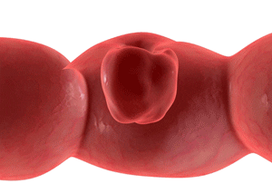
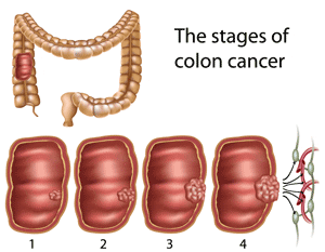
Colorectal Polyps
There are various types of polyps that can be found in the large bowel. The most common type of colon polyp is the adenoma which is a benign tumor (premalignant) that has the potential to develop into an invasive cancer if left in place and allowed to grow for years. Very rarely, polyps are found that have already developed into small cancers; these are called polyp cancers. Some polyps have begun the transformation to cancer; in this situation, microscopically, the polyp cells more closely resemble a cancer than an adenoma yet there is no invasion beyond the innermost layer of the colon wall. These are given the name dysplastic polyps or “carcinoma in situ”.
The second most common type of polyp, usually quite small, is called a hyperplastic polyp. Although hyperplastic polyps arise in the colon they are more often found in the rectum. Thankfully, hyperplastic polyps are not capable of transforming into a cancer. A third category of polyps, inflammatory, are found in patients who have had colitis related to inflammatory bowel disease, or infection. Thankfully, these polyps do not have the capacity to turn into a cancer.
Polyps come in a variety of shapes and sizes. Some are flat and grow directly on the surface of the colon while others are like small mushrooms that protrude into the colon on thin flexible stalks. Adenomas range in size from several millimeters (1/16 of an inch) to over 10 cm (3 inches). The chances that a given adenoma polyp may contain an invasive cancer is related to the size of polyp and to the type of adenoma that is present.
It is usually not possible to distinguish between adenomas (which can turn into a cancer) and the other types of polyps just by looking at them. Therefore, when polyps are found they are removed or at least biopsied. Most polyps are small enough that they can be removed or destroyed at the time of colonoscopy. A small percentage of polyps are too large to be removed with the colonoscope. These larger polyps are removed in the operating room by resecting a portion or segment of the colon or through a combined laparoscopic and colonoscopic method.
Treatment
The great majority of benign colon and rectal polyps are small or moderate sized and can be removed through the colonoscope in the outpatient setting using a variety of instruments that are passed through a narrow channel in the colonoscope. Very small polyps are destroyed with a forceps that grasps and removes small pieces of the rectal lining. Larger polyps are removed most often with a metal snare (like a noose) that is passed through a thin insulated hollow plastic tube. The snare and plastic sheath are passed through a channel in the colonoscope to the scopes tip and beyond. The snare is placed around the polyp (often shaped like a mushroom) and tightened while electric current is passed through the wire. This cauterizes the stalk of the polyp while it is being cut. If possible, the polyp is recovered and sent for pathological analysis. Polyps can also be directly burned or cauterized with heat or electric current, usually after one or several biopsies have been obtained. Larger polyps, especially the flat ones, are more difficult to treat colonoscopically.
Ideally, a polyp is removed completely and intact with a single application of the wire snare. However, this is not possible for some middle sized and larger polyps. One approach to these lesions is to remove them in pieces using the wire snare multiple times. To make it easier and safer to remove these larger polyps saline or other solutions are sometimes injected into the bowel wall via a catheter (with a needle at its tip) which is passed through the colonoscope. The injected fluid expands and swells the bowel wall making it thicker which protects the deeper muscle layers of the bowel wall from injury when the snare is used to remove parts of the polyp. In some cases it is not possible to fully remove the polyp at one colonoscopy. In this situation it is common to mark or “tattoo” the location of the polyp with India ink or another dye so that the area can be easily found. The colonoscopy is usually repeated months later at which time an attempt is made to remove the remaining polyp, usually with a metal snare, as before. It may take 3 or 4 colonoscopies to fully destroy a larger polyp using the colonoscope.
Unfortunately, when some polyps are initially found they are judged too large to be removed with a colonoscope in an endoscopy suite. Also, in some patients, after several attempts to colonoscopically remove a polyp, the gastroenterologist will make the judgement that the polyp can be fully destroyed with the scope. In these situations the patient is usually referred to a surgeon after the polyp has been tattooed with India ink.
Surgical Treatment of Large Benign Polyps
Historically, a segmental “cancer type” bowel resection is carried out to remove adenomas of the colon that are judged not amenable to removal with a colonoscope. In this case a 7 to 10 inch length of colon is resected (the polyp is usually in the middle of the specimen) along with the lymph nodes and blood vessels supplying the bowel after which the remaining ends are rejoined. Many patients ask why so much bowel is removed to treat a benign polyp? The reason is that 10 to 15 percent of large, supposedly benign, polyps that come to surgical resection are found to contain invasive cancers. Certainly, for the 10-15 percent of patients with cancers the lengthier and more extensive resection is logical and appropriate. In regards to the remaining 85-90 percent of patients with benign polyps the cancer type resection is not necessary. If there were a way to be reasonably certain that a polyp was, in fact, benign, then, perhaps, the radical resection could be avoided.
An important method of judging a polyp is by taking multiple biopsies of it through the colonoscope. Another useful method is to inject saline into the bowel wall beneath the polyp to see if the polyp “lifts” off the deeper layers of the bowel wall. The injected fluid greatly expands the middle layer of the bowel wall that separates the inner lining from the outer muscle coating of the colon. If the polyp rises, then the lesion is not invading into the muscular layer (a characteristic of invasive cancers). This saline lift test is easily done through the colonoscope. Yet another method is endoscopic ultrasound which uses sound waves to determine whether the polyp is invading and into the deeper bowel wall layers. Simply taking a close look at a polyp can also provide important information to the surgeon. What are the alternatives to a full segmental cancer type resection for polyps judged benign by the above tests?
Some large benign polyps can be removed by resecting a small oval shaped piece of the bowel wall (part of the circumference only) that includes the polyp and a small rim of normal bowel wall. This is called a "wedge" resection. This operation avoids extensive dissection and does not include division of the blood vessels supplying the area or removal of the lymph nodes. In short, it is a much smaller and less radical operation that requires minimal dissection of the colon and removes far less tissue. The chances of having a complication after this type of surgery is lower because less has been done. Patients usually go home in 1 to 2 days as opposed to 3 to 5 days after the standard cancer type bowel resection.
The “wedge” resection is best performed laparoscopically. Using this method the polyp and adjacent bowel wall is resected with a narrow stapler that is inserted through a hollow 1 inch "port" in the abdominal wall. The specimen is removed through one of the laparoscopic port wounds in a plastic bag after which a pathologist immediately examines the polyp and carries out one or several "frozen sections" to verify that the lesion is an adenoma only. The patient remains on the operating room table asleep while the polyp is evaluated. In the unlikely situation that the frozen section reveals an invasive cancer, then a standard cancer type resection would be immediately carried out laparoscopically.
Another way to remove some of these polyps is to perform a colonoscopy in conjunction with laparoscopy in the operating room with the patient under general anesthesia. The laparoscopic instruments can be used to push on the outside of the colon wall to make it easier for the doctor driving the colonoscope to grasp the polyp with a snare. In this way, some polyps that could not be removed during a regular outpatient colonoscopy can be excised. It is also possible to use advanced colonoscopic polypectomy methods to remove these polyps. One such method is called ESD or Endoscopic Submucosal Dissection. In this method a thin wire connected to an electric cautery machine is passed through an insulated sheath through the colonoscope and used to make an incision around the polyp (like a knife). Then other colonoscopic tools are used to lift and dissect beneath the polyp in order to fully detach it. If successful, at the end, the polyp has been removed in one piece and the underlying muscle layer remains intact. If successful, this method avoids removing even a “wedge” of the entire bowel wall.
Dr. Whelan’s Approach at Lenox Hill Hospital
My approach is to assess all patients with large benign polyps who are sent for a standard colon resection and determine if either the laparoscopic "wedge" or the combined laparoscopic / colonoscopic polypectomy methods can be utilized. The goal is to remove the polyp via the least invasive method possible. If successful, patients are home sooner with all or more of their colon in tact. Because it is not possible to be certain before surgery that a polyp can be removed via a combined laparoscopic/colonoscopic or wedge method, patients must consent to a standard colectomy in addition to the less invasive polyp removal methods. The consent states that, at the end of the procedure, the polyp will have been removed by one of the 3 methods with the standard cancer type resection being the last resort.
When the colonoscopy is performed in the operating room on the day of surgery if the polyp lifts when injected and is judged resectable via colonoscope then an attempt will be made to excise it using a variety of colonoscopic tools. If needed, the polyp can be manipulated externally with laparoscopic instruments to facilitate removal. If the polyp is successfully removed a test is done to make sure that the colon wall is not perforated after which the scopes are removed and the patient woken up.
If it is not possible to fully remove the polyp using colonoscopic methods, the borders of the polyp are marked with india ink and then an attempt is made to do a laparoscopic wedge resection of the polyp and the adjacent colon wall with a stapler. The colonoscope, still in place, views the placement of the stapler to male sure all is well. If successful, then the specimen is put in a plastic bag and removed through one of the small wounds. However, if the polyp will not lift when injected, is judged too large for colonoscopic or wedge resection, or looks like a cancer then the standard cancer type bowel resection would be immediately performed.
Colorectal Cancer Section
Introduction
Colon and rectal cancer is the 2nd or 3rd most common cause of cancer-related death in the United States. In the U.S. about 150,000 new cases are diagnosed each year. The colon and rectum are the 2 parts of the large intestine (or large bowel). The rectum is the last 5 inches of the large bowel that leads to the anus. The colon is about 3 to 4 feet in length and runs from the small intestine to the rectum. The vast majority of colorectal cancers start as benign polyps that develop from the inner lining of the large bowel that is called the mucosa. Although the exact time varies from patient to patient it is thought that, on average, it takes about 5 to 7 years for a polyp to develop into an invasive cancer. Periodic colonoscopy examinations are advised in order to detect and destroy benign polyps before they have the opportunity to develop into a cancer (see colonoscopy section). Colonoscopy is the most common way cancers are diagnosed.
Colorectal cancer can cause a variety of symptoms including bleeding, anemia, weight loss, a change in bowel habits or in the pattern and type of bowel movements and, rarely, pain or abdominal distension or bloating. In the U.S. a large proportion of large bowel cancers are detected on screening colonoscopy in patients who do not have any symptoms at all. Other ways that these tumors may be diagnosed are CT scans, PET scans, MRI scans, barium enema or virtual colonoscopy. Rectal cancers that are close to the anus are often discovered on digital examination of the anus which is a routine part of the physical examination carried out by general medical doctors or internists. Some patients are first found to be anemic (low red blood cell count) which leads to a search for a source of the blood loss. A colon exam is part of the evaluation for anemia. The best way to verify the presence of a cancer is to biopsy the tumor. This is most often done via colonoscopy.
After a large bowel cancer has been discovered other tests are usually carried out to determine if the tumor has spread to another part of the body. The most common places for a large bowel malignancy to travel (from the most to the least frequent) are the liver, lungs, bone, and the brain. Abdominal and pelvic CT scans, MRI scans, PET scans, or ultrasound examinations are the tests that are most commonly used (usually only 1 test is performed per patient). Also some blood evaluations are usually done including a CEA test (carcinoembryonic antigen). CEA is a cancer marker that most, but not all, colorectal cancers make and that can be found in the blood stream. Rectal cancer, for a number of reasons, is different from colon cancer in regards to the preoperative evaluation prior to treatment (please see the rectal cancer section). The treatment options for colon and rectal cancers will be discussed separately.
Colon Cancer Treatment
Surgical removal of the segment of colon that contains the tumor is the primary treatment for the great majority of colon cancers. Patients with Stage 4 disease are an exception and are usually best treated with chemotherapy initially. In patients that undergo surgery, the segment of colon removed is usually 7-10 inches long; in addition to the tumor the lymph nodes and blood vessels that supply the tumor are removed. In some patients with multiple lesions or a predisposition to colon cancer, the majority of the colon (2-3 feet) is removed. In the vast majority of colon cancer resection patients the remaining ends of the bowel are reconnected (anastomosis) so that the patient will go to the bathroom in the usual way (through the anus). In some patients a colostomy or ileostomy (aka stoma) may be necessary but is usually temporary; in this situation the end of the bowel closest to the stomach is brought out through the abdominal wall of the patient and sewn to the skin. A bag or appliance is placed over the stoma to catch the stool which exits the body through this opening rather than via the anus.
Presently, there are several surgical methods that can be used to carry out the colon resection (aka colectomy). Up until the early 90’s the only method available was the “open” method in which bowel resections were carried out through a lengthy incision made in the abdominal wall that provided the surgeon access to the abdominal cavity. The incision is usually a vertical one made in the middle of the abdomen although sometimes a side to side or transverse incision is used. The second method that is now available is called the laparoscopic or minimally invasive method. This method utilizes 4 or 5 small incisions (3/8” to 3/4” in size) through which hollow cylinders called ports are placed. Carbon dioxide gas is pumped into the abdomen through a port and elevates the abdominal wall creating a space within which the surgery is carried out. A long slender telescope with a camera attached is inserted into the abdomen and the operation is done with long thin instruments placed through the other ports. The proven advantages of laparoscopic methods are: 1) less pain, 2) quicker return of bowel function and resumption of oral diet, 3) more rapid discharge home, and 4) better preserved immune function. Please see the Minimally Invasive Surgery Section for a more detailed explanation.
After surgery, some patients will be advised to take chemotherapy, usually for a period of 6 months, in order to improve their chances of avoiding a cancer recurrence and to improve their chances of survival. Chemotherapy is recommended for patients who on review of the colon specimen are found to have 1 or more lymph nodes involved with cancer and for some patients with tumors that invade through the entire colon wall. Radiation treatment, except in very rare situations, is not given to colon cancer patients. Currently, there are at least 6 chemotherapy drugs which have been proven to be effective for colorectal cancer. A variety of regimens are being used today (please visit the Continuum Cancer Center of New York’s Medical Oncology Site for more details). As mentioned above, most patients found to have metastatic colorectal cancer, with the exception of those whose large bowel is nearly obstructed and those who are bleeding heavily, are initially treated with chemotherapy alone. In these cases, surgery is held in reserve.
Rectal Cancer Evaluation and Preoperative Radiation and Chemotherapy Treatment
The rectum, the part of the large bowel leading to the anus, is about 5 inches long. Cancers in this segment are treated differently than colon tumors. Unlike the colon, two thirds of the rectum is embedded in the pelvic tissue and is situated very close to the vagina and bladder in women and to the prostate and bladder in men. Rectal cancer is harder to cure, in part, because it can more readily spread to these surrounding pelvic organs as well as to the sacrum bone. Also, for rectal cancers that involve the anus or are located very close to the anus it is sometimes necessary to surgically remove the entire anus and rectum and make a permanent colostomy (end of the remaining colon brought out through the abdominal wall). Radiation (RT) and chemotherapy (chemo) are often given before surgery in an effort to improve survival, reduce recurrence rates, and save the anus so that it can then be re-hooked up to the colon. RT and chemo can also be given after surgery, however, it has been proven that it is more effective when given before surgery.
Studies of rectal cancer patients have shown that preoperative (preop) RT and Chemo increases the chances of saving the anal sphincter and also significantly decreases the chances that the cancer will come back in the pelvis. Preo RT and Chemo can also convert an unresectable tumor into a resectable one. Not all patients are advised to get preop RT and chemo; only patients whose tumors invade through the entire rectal wall into the surrounding tissue OR who have enlarged lymph nodes in this area are given this pre-surgical treatment. In order to determine how far the tumor has invaded and whether there are enlarged lymph nodes one or several tests are done after the rectal cancer has been diagnosed.
The 2 best ways to evaluate rectal cancers are via ultrasound (transrectal ultrasound, TRUS) and magnetic resonance imaging (MRI). The ultrasound examination is done by inserting either a rigid or flexible ultrasound probe into the rectum via the anus and then examining the cancer. The rigid probe is most commonly used in the U. S. . The depth of invasion of the cancer can be determined accurately and enlarged lymph nodes can also be detected. Tumors that invade through the entire wall into the surrounding fatty tissue are called T-3 lesions (T-1 and T-2 lesions invade only partway through the rectal wall). The TRUS exam usually takes 15 to 30 minutes to complete and is well tolerated by most patients. The MRI scan is done with a large doughnut shaped machine into which the patient is positioned. In addition, for a detailed pelvic examination, a special blanket is usually placed over the patient’s lower abdomen and pelvis. The MRI provides very detailed information including the depth of invasion and the presence of enlarged lymph nodes. Dr. Whelan’s office is equipped with an ultrasound machine and, routinely, rectal cancer patients undergo a TRUS exam at the time of their first visit (limited bowel preparation required). The St. Luke’s Roosevelt Hospital and CCCNY have several state of the art MRI machines at several locations. Although MRI exams are offered at many locations in the metropolitan area, for patients who decide to undergo treatment with Dr. Whelan it is advised that the MRI be done at a CCCNY location so that the results and images will be readily available to the team of doctors involved in the care of the patient.
In addition to the TRUS or MRI done to locally evaluate the cancer, these patients are also evaluated for distant metastases (liver, lung, etc) via CT scans, MRI scans, PET scans, and/or ultrasound examinations, as mentioned earlier.
Surgical Treatment Options For Rectal Cancer
Similar to colon cancer, surgery remains the mainstay of curative treatment for rectal cancer. Presently, world wide, the great majority of rectal cancers that have not spread to distant sites (liver, lung, etc) are surgically resected through the abdomen using either open (big incision) or laparoscopic methods. A small percentage of rectal cancers are resected through the anus (local excision method) at some hospital centers. These are usually superficial lesions located close to the anus yet above the anal sphincter that do not invade through the full thickness of the rectal wall.
Local Surgical Treatment of Rectal Cancer
As mentioned, transanal (through the anus) local excision is an option for select rectal cancers. In this operation a “disc” of the rectal wall containing the cancer and a margin of the normal rectal wall is resected through the anus. The resulting wound is usually, but not always, closed with sutures. Possible complications include bleeding and infection. This is a far less traumatic operation than the “through the abdomen” radical rectal resection method. Local excisions are usually done with a spinal anesthetic which means that general anesthesia can usually be avoided. One major downside to this method is that it does not remove the lymph nodes that supply the rectum; if the tumor has spread to the lymph nodes then the patient is very likely to develop a tumor recurrence unless radiation and chemotherapy are given. In contrast, the through the abdomen “radical” resection does remove the lymph nodes in the area. Because of this major drawback, the local excision approach is usually restricted to consenting patients with superficial cancers who have only a 5 to 15 percent of lymph node involvement. It is usually combined with radiation and chemotherapy in order to treat the lymph nodes in the pelvis.
The complication rate for the local excision method is low and patients are usually discharged home within a few days. Unfortunately, local transanal rectal cancer resection is associated with significantly higher local cancer recurrence rates than the radical, through the abdomen, rectal resection. As of 2010, the “gold standard” for curative rectal cancer surgical treatment remains the through the abdomen approach.
In addition to transanal local excision, alternative local treatments for rectal cancer include coagulating or burning the tumor (aka fulguration) or high dose radiotherapy that is applied directly to the tumor through a scope placed through the anus. The local excision method is the most commonly employed local treatment option. As of 2010, as mentioned above, local excision is usually combined with preoperative RT and Chemo in an effort to decrease the local recurrence rate. Early results using this approach are promising. Proponents of the RT and Chemo followed by local excision are in the midst of organizing a multicenter trial that they hope will provide data demonstrating that, for select cancers, this approach yields results comparable to those following radical through the abdomen rectal resection.
Through the Abdomen “Radical” Rectal Resection for Rectal Cancer
Trans-abdominal rectal resection remains the treatment of choice and is associated with the highest rate of survival and lowest recurrence rates. In this operation the tumor along with a margin of normal rectum (and usually some colon as well) is removed in addition to the lymph nodes and blood vessels supplying the rectum. The rectal resection method that is the “gold standard” routinely employed in the U.S. and worldwide is called ‘total mesoretal excision’ or TME technique. Operations performed using the TME method when carried out by experienced surgeons yield the lowest local recurrence rates and the highest 5 year survival rates.
Essentially there are 3 different radical operations for rectal cancer (TME method is used for all). Which operation is performed depends mainly on how far away from the anus and anal sphincter the tumor is located. In order to cure the patient it is necessary that a cuff of normal rectum (known as a “margin”) beyond the tumor (closer to the anus) be excised to ensure that the entire tumor has been removed. As long as there is some rectum left in the patient it is usually possible to reconnect to remaining colon to the rectal remnant (the rejoining is called an anastomosis) so that the patient will be able to continue going to the bathroom in the usual way. This operation is called a Low Anterior Resection (LAR).
If the anus is involved with the cancer then it is usually necessary to fully resect the anus, the anal sphincter muscles, and the rectum to safely remove the tumor. In this situation the end of the remaining colon is brought out through a circular opening in the abdominal wall to create a permanent colostomy. Stool then exits the body through the colostomy into an airtight plastic bag that is securely attached to the surrounding skin. This operation is called an AbdominoPerineal Resection (APR).
If the tumor is located just above the anus and sphincter such that it is not possible to resect a cuff of normal rectum, in some cases, it may be possible to remove the last inch of the rectal lining while preserving the anal sphincter muscle. The remaining colon can then be sewn to the anal skin in order to permit the patient to go to the bathroom through the anus in the normal way. This operation is called a proctectomy with a coloanal anastomosis (Coloanal).
One important complication that can occur during the first 10 days after surgery in LAR or Coloanal cases is an anastomotic leak. In this situation small amounts of stool leak out between the sutures or staples of the anastomosis into the surrounding pelvis and usually cause an abscess. Patients with leaks usually get quite ill and often require reoperation to drain the abscess and to bring either the colon or the small bowel outside the body through a circular opening in the abdominal wall. The externalized bowel is opened and sewn to the skin edge to create either an ileostomy (small bowel) or colostomy (colon) that redirects or “diverts” the stool to the abdominal wall. This stops the flow of stool to the anus as well as the seepage from the anastomosis and, in most cases, allows the leak to seal. Patients who develop anastomotic leaks are usually in the hospital for at least 2 weeks if not longer. Once the leak seals and the infection fully resolves the ileostomy or colostomy can be closed at a smaller second operation. After closure the patient will go to the bathroom through the anus in the usual way. Unfortunately, bowel function after an anastomotic leak is usually worse (more bowel movements and a decreased ability to store stool) than in patients who heal uneventfully.
In an effort to avoid anastomotic leaks surgeons often make a temporary ileostomy or colostomy (aka ‘stoma’) at the time of the rectal resection and anastomosis. If, from the start, the patient has a stoma that diverts the stool away from the bowel rejoining point then it is highly unlikely that a leak, abscess or pelvic infection will develop. The average length of stay in patients with a stoma after an LAR or Coloanal operation is about 6-9 days. The temporary stoma is closed at a smaller second operation (usually less than one hour) that is usually carried out about 2-3 months later after verifying that the anastomosis has healed properly.
All 3 radical rectal resection operations (LAR, APR, and Coloanal) can be carried out using either open (big incision) or minimally invasive surgery (MIS) methods. MIS operations are associated with significantly smaller abdominal wall incisions as well as less pain, a quicker bowel recovery, and a shorter hospitalization. Please see the Surgery Technique Section for more information about both MIS and Open operations. Dr. Whelan uses MIS methods for 85 % of his rectal cancer operations and has done so for the last 14-15 years.
Colostomy/Ileostomy
There are times during colon surgery that it may be necessary for the surgeon to create an ostomy. An ostomy is a surgically created opening, usually in the abdominal wall for the discharge of body wastes. The intestines are “re routed” so that instead of the stool passing through the colon and out the rectum, there is and opening for effluent to pass through the abdominal wall and in to an external pouch. The opening that is seen on the abdominal wall is called a stoma.
Ostomies can also involve any part of the small or large intestine. They can also, in the case of urinary bladder cancers, be formed to collect urine.
The portion of the intestine that is brought out to the abdomen determines whether or not the patient has a colostomy or an ileostomy. A colostomy is created when a portion of the colon is brought out to abdominal wall. The term ileostomy is used when a portion of the small intestine is used to form the stoma. Colostomy out put is generally more formed and regular, whereas that of an ileostomy is more frequent and semiformed.
Ostomies can be either temporary or permanent, depending on the situation. Temporary ostomies are utilized when the surgeon is concerned with the possiblilty of an anastomotic leak forming post operatively (see section on colon cancer/diverticulitis). In this case the stool is temporarily diverted in order to give the anastomosis (internal suture line) a chance to heal. The temporary Ostomy is generally reversed, after testing, approximately 3 months after the initial surgery. Permanent ostomies may be necessary when there is a low rectal cancer, or when the patient cannot withstand a second surgery for closure.
The idea of having an ostomy can be a very emotional consideration for many patients. It is important to understand that ostomates (persons who have ostomies) can and do live normal lives. Once over the recuperative phase of surgery most people with ostomies do everything they were able to do before. There is a learning curve that has to be met before feeling secure, but taking care of an ostomy is not hard and there is no one right way to manage it. It is important to remember that the ostomy must fit in to your life; you do not change your life to revolve around the ostomy. Once learning the basics, most people go on to figure out what works best for them.
No one should refuse potentially life saving surgery simply because they do not want to “wear a bag”. In fact, many times the creation of a colostomy or ileostomy can actually improve the quality of a patient’s life. This is particularly true in someone with a history of severe colitis or fecal incontinence where planning daily activities must always include knowing where the nearest bathroom facilities are.
There are many different types of appliances/pouches available and Wound, Ostomy & Continence (WOCN) nurses can help with the selection. WOCN’s are registered nurses who specialize in the care of patients who have undergone ostomy surgery. Having access to a WOCN is crucial pre and post operatively. WOCN nurses can help with stoma placement pre op so that the ostomy does not interfere with clothing and is also critical in helping with appliance selection post operatively
Appliances are like any other type of clothing or prosthesis, they must be fitted to the patient to ensure optimum success. Sometimes initially after surgery, when there are still sutures and abdominal swelling, there may be a temporary problem with fit but that should never be accepted as “normal”.
The United Ostomy Association of America and the Wound, Ostomy & Continence Nurses Society are tow organizations who provide support and education for ostomy patients.
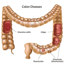
Diverticulitis
Introduction: Colonic diverticuli are small pouches or sacs that protrude outwards from the colon wall. Although they can be found in any part of the colon by far and away the most commonly involved segment is the sigmoid colon. It is believed that the great majority of diverticuli are acquired, meaning that they develop during the course of a person’s life. Although no one is certain, most experts believe that diverticuli form as a result of high pressure in the colon that develops when the large bowel contracts. The colon moves stool towards the anus by periodically contracting in a sequential fashion. These contractions temporarily raise the pressure in the colon. In some patients the pressures that develop can be quite high and it is thought that over the course of years the outpouchings slowly develop. Some experts believe the typical low fiber Western diet results in hard dense stools and contributes to the development of diverticuli since the colon must generate higher pressures to move hard stool through the bowel. The rate of diverticulosis in non-vegetarians is almost 3 times higher than in vegetarians. It is estimated that at least 30 % of Americans over the age of 60 and perhaps 60 % of those over 80 have “diverticulosis” which is the name given to patients who have diverticuli in one or more parts of the colon.
Acute Diverticulitis: In some patients with diverticulosis the colon wall becomes inflamed and a small perforation or hole may develop in a diverticulum that leads to an infection in the abdomen called “diverticulitis”. The most common symptoms of acute diverticulitis are lower left abdominal pain and fever. Other symptoms that may develop include a change in bowel habits, swelling of the abdomen, and nausea and vomiting. Currently, the diagnosis is almost always made with a CT scan of the abdomen which shows characteristic signs of thickening and inflammation of the colon wall. Attacks of diverticulitis can be classified as simple or complex. Thankfully, the majority are simple attacks that can be treated with antibiotics and restriction of diet alone. These episodes most often resolve within a week although some may require several weeks of antibiotics. In most patients the abdominal pain and other symptoms are much improved within 48 hours. The majority of patients with simple attacks are treated as outpatients with oral antibiotics. Some patients with simple attacks require hospitalization and intravenous antibiotics. These patients are usually discharged once the fever and abdominal pain has resolved and are continued on oral antibiotics for at least a week.
Complex Diverticulitis: Unfortunately, some patients have complex attacks of diverticulitis that are more difficult to treat; these patients all require hospitalization. For example, an abscess can develop next to the inflamed colon that may not go away with antibiotics alone. Large abscesses are most often treated with a drainage tube that is placed through the abdominal wall or buttocks by a radiologist. Typically, the tube stays in for a week or longer. In most cases an emergency operation can be avoided, however, most surgeons advise an elective colon resection after resolution of the acute abscess and diverticulitis in order to prevent future problems. Very rarely, an urgent operation is needed to drain an abscess and, possibly, to remove the diseased bowel and/or make a temporary colostomy. A very small number of patients with diverticulitis develop diffuse peritonitis with severe widespread abdominal pain, high fever, and other signs of severe illness. These patients almost always require immediate surgery. Another complex problem that can arise in patients with diverticulitis is an abnormal connection or fistula between the colon and the urinary bladder, another piece of intestine, the vagina, or the abdominal wall. The most common type is a colon to bladder fistula (colovesical fistula) that is usually discovered when patients note they are passing a combination of gas and urine when urinating. Patients with colovesical fistulas may also develop difficult to treat and frequent urinary infections. Patients with a diverticular fistula require an elective colon resection to resolve this problem. Rarely, patients with acute diverticulitis may develop a complete obstruction that will require surgery to resolve (see Other Diverticular Disease section).
There is a group of patients with acute diverticulitis without a sizable abscess, fistula, diffuse peritonitis, or obstruction whose attacks do not readily resolve. These patients may have persistent symptoms or develop recurrent symptoms soon after completing the prescribed course of antibiotics. In some cases it may take months of treatment with different antibiotics to get the patient over the episode. These attacks are complex from the standpoint of being persistent. Some of these patients may have small abscesses that are judged too small to drain through the skin. Other patients, in addition to persistent symptoms, are repeatedly found to have air outside of the colon wall on CT scan (a sign of perforation). These patients, because of their complex first attack, are more likely to have future attacks and may be best off having an elective colon resection without waiting for a second or third attack to develop.
Colon Screening After Diverticulitis: Patients with diverticulitis who have never had a colonoscopy or other colon screening examination need to have a colonoscopy done a few months after their first attack to rule out colon tumors. Rarely, colon cancers can be confused with diverticulitis on CT scans. The colon evaluation is not done during or immediately after an attack because of fear that the examination may re-activate the diverticulitis. The alternative screening methods are barium enema and virtual CT scan.
When is Surgery Necessary? Surgery is not advised for patients who have had a single attack of simple diverticulitis. In these patients no further attacks occur in between 40 and 70 percent of patients. These patients should undergo standard colonoscopy for colorectal polyps and cancer at the standard intervals. The risk of future attacks of diverticulitis increases notably in patients who have had 2 discrete attacks. Most patients who have multiple attacks of diverticulitis have attacks similar to their first, however, in a small percentage a more complex attack may develop. It is very important that patients undergo CT scans when they have their second, third, or later attack of diverticulitis to confirm the diagnosis and also to make sure that an abscess has not developed. Most experts agree that elective surgery to remove the involved colon is reasonable after 2 episodes. After 3 clear cut episodes it can be confidently predicted that future attacks will develop and for this reason surgery is recommended.
One factor that should be taken into account when making a decision about surgery is the length of time between attacks. If the interval is 3 to 5 years and the attacks have been mild and easy to treat then a patient may decide to avoid surgery and to take their chances that there will be few future episodes. If, however, the interval between attacks is 3-6 months or is shortening with each attack then a very strong case can be made that surgery is the best course of action. Another factor that should be considered is the nature of recurrent attacks. Patients whose attacks become more difficult to treat or who have urinary symptoms (urinary frequency or painful urination) run a higher risk that an abscess or fistula may develop.
Operations for Diverticulitis: A basic principle of surgery for diverticulitis is that the chances of needing a temporary colostomy or ileostomy is much lower when a colorectal resection is done electively a time when the patient is not having in the midst of an attack. Surgery is more difficult in the setting of acute inflammation and an active infection.
Elective Operations: The ideal operation for a patient with diverticulitis is one where the diseased segment of colon is resected and the remaining ends of the large bowel are rejoined together without a temporary colostomy or ileostomy. This operation is possible for the great majority of patients who have an elective colon resection between attacks. However, in about 5 to 8 percent of elective resection patients, although it is possible to reconnect the 2 ends of the bowel, a temporary stoma (ileostomy or colostomy) is made nonetheless to divert the stool away from the new anastomosis. The protective stoma is helpful in this situation because it greatly decreases the chances of an anastomotic leak or abscess developing by preventing the stool from flowing past the rejoining point.
In a very small percentage of patients, after the bowel resection has been completed, the two ends of the large bowel are not rejoined and a colostomy is made instead. One reason for this decision may be the presence of severe inflammation or scarring that makes it difficult to construct the anastomsosis. Another reason may be that the end of the remaining colon may be too short to reach the rectum. Yet another reason may be that the time required to perform the resection was so long that the surgeon and anesthesiologist decide that the bowel rejoining should be put off until another day so that the operation is completed sooner. A final reason for not rejoining the bowel ends is if the patient is unstable from the anesthesiologists point of view in regards to cardiac, pulmonary, or other reasons. A second separate operation to “take down” the colostomy and rejoin the bowel ends can be done at a later date for some of these patients.
In an exceedingly small percentage of patients (less than 1 %) who undergo elective surgery it may not be possible to resect and remove the diseased part of the colon because of severe disease. In these cases the surgeon may not be able to identify the tissue planes between the sigmoid colon and the other organs of the pelvis and lower abdomen. Whereas in most cases a safe approach can be found, in this very small subgroup, resection is not feasible without risking major injury to the bladder or the major blood vessels and other important structures of the pelvis. The only option in this situation is to make a colostomy to keep the stool away from the diseased part of the colon. In these patients it is usually possible to resect the diseased bowel and reconnect the 2 ends at a second operation done 6 to 12 months after the first procedure.
Emergency Operations: In very rare situations it is necessary to do surgery during an acute attack of diverticulitis. The most common reasons for this are: 1) diffuse peritonitis, 2) an abscess that cannot be drained with a tube placed through the abdominal wall, 3) failure to improve on intravenous antibiotics and 4) complete colonic obstruction. In the emergency setting the surgery is usually more difficult which increases substantially the chances that a stoma (colostomy) will be needed. If possible the diseased piece of colon is resected and the two ends rejoined. Even when this is possible a temporary stoma (ileostomy) is usually made to protect the newly rejoined colon and rectum. It is more common in the emergency setting for the surgeon to do the colon resection and to make a stoma without performing an anastomosis. Most emergency surgery patients who have a colostomy can have the stoma removed and the large bowel ends re-connected at a second operation that can be safely done 3 to 6 months later.
Surgical Method and Approach: The great majority of elective operations for diverticulitis can be carried out laparoscopically. The advantages of minimally invasive operations include less pain, earlier bowel function recovery and earlier discharge from the hospital. The choice of surgical method, open or laparoscopic, does not influence the ability of the surgeon to do the bowel resection. In fact, the laparoscopic approach facilitates the mobilization of the splenic flexure portion of the colon which is required in the majority of patients in order to safely rejoin the two bowel ends. If severe inflammation is found in the pelvis and it proves necessary to convert a laparoscopic case to open surgical methods the patient still benefits as long as the part of the operation that is outside the pelvis is completed laparoscopically. In this situation the final incision is usually about 11 cm long as opposed to the incision required to perform the operation using fully open methods (on average 22-26 cm long). This use of both laparoscopic and open methods is called the hybrid approach. In a small percentage of patients with severe disease a fully open operation may be the best choice.
Other Diverticular Disease
Bleeding: A small percentage of diverticulosis patients develop heavy rectal bleeding. For reasons that are unknown, major bleeding does not occur during an attack of diverticulitis. Although most bleeding episodes stop spontaneously some patients bleed repeatedly and require blood transfusions or other treatment. Because bleeding can occur in any part of the colon, when patients develop heavy rectal bleeding doctors must determine the location and, if possible, the cause of the hemorrhage. There are several ways to evaluate patients with severe bleeding. Colonoscopy is one method that can be used to locate the bleeding site. Another method, called angiography, involves injection of contrast into the bloodstream after which X-rays movies of the blood vessels that supply the colon are carried out. If the bleeding is active and heavy enough the contrast can be seen “pooling” in the colon at the bleeding site on the x-ray. These x-rays reveal which part of the colon is bleeding. The final diagnostic option is a nuclear bleeding scan. In this test a radioactive “marker” is intravenously injected after which a body scanner is used to look for pooling of the contrast in the abdomen. If the patient is bleeding at the time of the scan and is heavy enough then pooling will be seen. The location of the pooled blood provides clues as to which part of the intestine is bleeding.
Patients who have multiple bleeding events or severe hemorrhage may require treatment. Surgical resection of a limited length of the colon will deal with the problem provided the bleeding segment has been identified preoperatively. In patients who require surgery in whom it is not possible to identify which part of the colon is bleeding, the entire colon or almost the whole colon is surgically removed. Depending on the patient’s condition, age, and other medical problems, the remaining bowel ends may be rejoined (anastomosis) or a temporary colostomy or ileostomy may be constructed. In the setting of heavy acute bleeding “open” large incision surgery is usually performed although in a minority of cases laparoscopic methods may be used.
There are 2 non-surgical treatment methods that can also be used to treat heavy gastrointestinal bleeding. Both methods involve angiography. In one, small coils or sponges are injected through the angiography catheter directly into the bleeding vessel in an effort to obstruct the vessel and stop the bleeding (called embolization). In the second method, after the angiography catheter has been inserted into the artery that is bleeding, medicines that cause constriction of blood vessels are injected directly with the hope that the bleeding will cease.
Obstruction: Diverticulitis usually results in thickening of the colon wall. In a small number of patients who have a history of multiple attacks the bowel wall gets so thick that it narrows the colon segment greatly. In some a complete obstruction can develop. Rarely, this type of obstruction develops in patients who have never had a recognized attack of diverticulitis. Patients with acute obstruction stop having bowel movements. Over the course of days, the abdomen becomes distended and swells. This condition is most often diagnosed with abdominal X-rays or a CT scan. Most patient with large bowel obstruction require urgent or emergent surgery at which time, ideally, the obstructed and narrowed segment of colon is removed. If possible, the remaining ends of the bowel are rejoined immediately. Unfortunately, in some patients this may not be possible or advisable; in this situation a temporary colostomy (colon) or ileostomy (small bowel) is constructed.
In some diverticulitis patients with obstruction it may not be possible to remove the diseased part of the colon at the initial operation. The most common reason is severe inflammation and adhesions between the diseased colon and the bladder or other pelvic tissues that make surgical dissection extremely difficult and hazardous. In these situations a colostomy is made which diverts the intestinal flow away from the obstructed segment and through the abdominal wall (see colostomy section). It is usually possible to do a second operation months later at which time the colostomy is closed so that the patient can go to the bathroom normally.
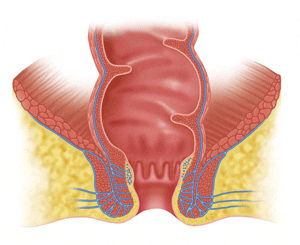
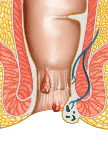
Hemorrhoids
Definition: Hemorrhoids are essentially dilated blood vessels, mostly veins, that develop over the course of years in response to having bowel movements. When humans defecate they sit down on the toilet and push after taking a breath and closing their airway (valsalva maneuver). The Valsalva maneuver increases the pressure inside the abdomen and pelvis which pushes the blood in the rectal veins into the anus. The veins fill with blood and, over the course of years, dilate. As the hemorrhoids develop the anal lining swells and protrudes into the anal canal. During BM’s the hemorrhoids get pushed towards the outside along with the stool. As they enlarge and develop, the inner or internal hemorrhoids may travel to the point where they are actually protruding outside from the anus. It is the pushing, swelling, and movement of the inner rectal lining that cause most of the symptoms associated with hemorrhoids. There are two types of hemorrhoids, internal and external. To understand the difference between internal and external hemorrhoids you must learn a little about the anatomy of the anal canal.
Anal Anatomy and Internal and External Hemorrhoids: The anal canal is about 1 to 2 inches in length and has 2 types of lining. The external lining is a specialized type of skin while the inner lining is rectal mucosa (pink in color and the type of lining found throughout the rectum and colon). The juncture of these two surface linings is in the middle of the anal canal (called the Dentate line). Hemorrhoids are classified as being either internal or external based on what type of lining lies over them. If there is skin covering the hemorrhoidal veins they are external and if there is pink rectal mucosa overlying them they are internal hemorrhoids. It is important to note that most hemorrhoids have both internal and external parts (called a mixed hemorrhoid). Having said this, there are patients who have either internal or external hemorrhoids alone. The symptoms patients experience depend very much on the type of hemorrhoids they have (internal, external, or mixed internal/external).
Thrombosed External Hemorrhoids: These very annoying and painful hemorrhoids tend to occur “out of the blue” and to last for 1-2 weeks only. In response to straining or having a BM the external hemorrhoid swells with blood. For some reason the veins that supply the external hemorrhoid do not allow the blood filled hemorrhoid to empty. It is believed that the vein thromboses (clots) which prevents blood flow. The arteries feeding the hemorrhoid continue to pump blood into the hemorrhoid which increases the swelling. Eventually, the veins burst and then the blood forms a clot. The localized swelling usually is purple and looks like a grape or marble has been slid under the skin. The thrombosed hemorrhoid stretches the anal skin overlying it which results in pain. The pain is constant and not related to BM’s. Having a BM does not make the pain worse or better. Eventually, if left alone, the hemorrhoid either spontaneously bursts, which relieves the pressure and lessens the pain, or is slowly resorbed which also decreases the swelling and the pain. Eventually, the swelling and the symptoms resolve. What is left in most patients is an asymptomatic skin tag. It is believed that external hemorrhoids cannot recur at the same location. Unfortunately, a new one can develop at another site around the anus.
Treatment of Thrombosed External Hemorrhoids: What is done for these painful hemorrhoids depends on when the patient is seen by the surgeon. If seen at a time when the pain is at its peak or increasing (usually within the first 3-5 days) then a simple surgical procedure, done in the office, is the best option. After administering a local anesthetic, a small incision is made over hemorrhoid and the blood clot removed. This relieves the pressure and the pain. The skin incision causes some pain afterwards, however, it is much less painful than the thrombosed hemorrhoid early in its course
If the patient is not seen by a doctor for a week or more after the onset of symptoms, most often, the symptoms have begun to decrease. The hemorrhoid may have spontaneously ruptured or started to resolve. The swelling may still be present and obvious, however, the pain is usually notably less and decreasing daily. If this is the situation then surgical incision should be avoided since, at this point, the pain from the surgical incision will likely be worse than the residual pain from the resolving hemorrhoid. Sitz baths, stool softeners, and non-steroidal analgesics such as Tylenol or advil are the best treatments at this point.
Hemorrhoid Classification: Internal Hemorrhoids are classified in regards to the extent to which they move and remain outside the anus (prolapse) during and after a bowel movement (BM). Grade 1 hemorrhoids do not appreciably prolapse during a BM. Grade 2 hemorrhoids briefly prolapse outside the anus but then immediately return to their regular position. Grade 3 hemorrhoids prolapse during a BM and have a tendency to remain outside the body for a period of time. If left alone they will eventually slide back into the rectum. Most people learn to push them back inside (“reduce”) with a finger or by sitting down on a hard surface after the BM. Grade 4 hemorrhoids remain prolapsed outside all of the time and will not stay inside when pushed back inside. When the inner rectal lining is continually exposed to the external environment it may become thicker and take on a different color. These “metaplastic” changes can be easily recognized by a colorectal surgeon and are not dangerous in regards to the development of a cancer.
Surgical Treatment of Hemorrhoids
Introduction: There are two basic surgical methods used to treat hemorrhoids: the standard hemorrhoid excision and the stapling method. Surgery is usually used only for Grade 3 or 4 hemorrhoids (hemorrhoids that prolapse outside the anus during a bowel movement and require a push or a long period of time before they go back inside.
Traditional Hemorrhoid Excision: This method is usually used for patients with mixed internal and external hemorrhoids. Each hemorrhoid group is removed (while preserving the anal sphincter and most of the anal lining) after which the wounds are closed with absorbable sutures. See below for more details of the procedure.
Hemorrhoidal Stapling: This method is best suited for patients with symptomatic internal hemorrhoids (most common symptoms are bleeding and prolapse) although it can also be used for patients who have large internal hemorrhoids and smaller external hemorrhoids. There is less pain associated with the stapling method and patients require less pain medications than traditional hemorrhoidectomy patients. With this method, part of the internal hemorrhoids and rectal lining close to the anal sphincter is removed with a staple gun that is inserted into the anal canal. There are no external incisions, sutures, or staples with this method. Hemorrhoidal stapling takes place in the operating room (same as the traditional hemorrhoidectomy method).
Holding Area / Consent: The patient will be admitted to the hospital in the Ambulatory Surgery area and an intravenous line placed. Either in the Ambulatory Surgery area or outside of the operating room both the surgeon and the anesthesiologist will speak with the patient, review the surgical and anesthesia options, and obtain consent from the patient. The patient will then be taken to the operating room.
Anesthesia Options:
Patient Positioning: The patient will be turned and placed face down lying on his stomach. A bolster (a pillow or rolled sheet) is placed under the patient’s pelvis to elevate the anorectal area. In addition two smaller bolsters are placed under the chest between the breast and the shoulder on each side. The buttocks are gently taped to the side of the table to expose the anorectal area. The anorectal area will be prepared with a soap solution to kill the bacteria on the skin. The anal canal and hemorrhoids will be examined after which the operation will be started.
Standard Excisional Approach: Usually there are 3 separate hemorrhoids to be dealt with (right anterior, right posterior, and left lateral). A solution of saline and dilute epinephrine is injected into the hemorrhoid before the dissection is begun in order to reduce the bleeding during the removal. The anoscope will be positioned and an inverted “V” shaped incision made at the outer edge of the hemorrhoid (see Figure 15-7). The first goal is to identify the anal sphincter muscle and to carefully dissect it away from the hemorrhoid. Once the muscle has been fully protected and the hemorrhoid fully elevated a thin oval shaped piece of the hemorrhoid is excised (see figure 15-8). The edges of wound and the remaining hemorrhoid are then elevated off of the muscle (to protect the muscle) which creates a flap after which the hemorrhoid tissue is excised from the flap while preserving the anal skin and the rectal surface lining (see figure 15-11). The hemorrhoids lie beneath the surface lining but above the anal muscle layer. The hemorrhoid can be excised without damaging either the anorectal lining or the muscle below it (this is called a submucosal resection). After the hemorrhoid has been fully excised, if there is bleeding it is stopped with hemocautery (electrical current used to coagulate the bleeding vessel) or a suture. Next the two edges of the wound are sutured together starting at the base of the wound (furthest inside the anal canal) and progressing towards the outer edge of the anus where the suture is tied. An absorbable running suture (over and over suture) material is used that does not need to be removed. The wound is fully closed in this manner (see Figure 15-13).
The same procedure will be carried out for each of the patient’s hemorrhoids. Usually, a total of 3 areas are resected (rarely 4). For the final 1 or 2 hemorrhoids, usually the smallest, a fully submucosal method may be used. In this method, after injecting the epinephrine solution, a linear incision is made along the long axis of the anal canal (about 4 to 6 cm in length). The sphincter muscle beneath is located and the hemorrhoid dissected away from the muscle (in order to protect the muscle). Next, the hemorrhoid is cut away from the flap but all of the surface lining is preserved. This is different from the above method in which an ellipse of anal lining is excised along with a portion of the hemorrhoid. After the hemorrhoid has been fully removed hemocautery is again used to stop any bleeding that may be present. The resulting wound is then suture closed using an absorbable suture.
At the end of the operation all suture lines are inspected and a local anesthetic is injected into the anal area for postoperative pain relief. A gauze dressing is then placed over the anus and held in place with mesh underwear or tape. The patient is then turned onto his back (from the face down position) and woken up by the anesthesiologist.
In this operation the anal muscles are fully protected and preserved. Also, great care is taken to ensure that the anal skin lining is adequately preserved. None of the nerves supplying and running to the anus are cut or injured in this operation
Postoperative Care
Three types of pain medication will be used: 1) flagyl, 2) toradol (ketorolac), and 3) vicodin (or percoset). Flagyl (metronidazole) is an antibiotic that has been shown to significantly decrease the amount of pain experienced by hemorrhoidectomy patients. The flagyl is taken for 5 days in total. A total of 3 flagyl pills are taken each day. Toradol is a very strong non-steroidal anti-inflammatory drug (NSAID) that is taken by mouth 4 times a day for 3 days. The toradol is an excellent pain medication for intestinal and anal operations because it is strong and doesn’t contain narcotic. Narcotics can be taken, however, they slow down intestinal motility and can lead to constipation. The last pain medication that is given, when the toradol and flagyl together are not providing adequate pain relief is vicodin. Vicodin is a mixture of narcotic and Tylenol that is very effective.
When the patient no longer requires narcotics (usually 3 to 5 days after surgery) but still wants some pain relief then ibuprofen (advil) 600 mg will be taken every 4 to 6 hours as needed. Ibuprofen does not cause constipation and is a very effective pain medication.
In addition to the above pain medications, the following medications are taken to soften the stool and prevent hard stools and constipation. Metamucil or any other phsyllium husk or cellulose based fiber additive (such as konsyl, or citrucel), is taken 1-2 tablespoon per day. The second medication that is taken is colace (docusate sodium) 100 mg, three times a day for 3 weeks. Finally, mineral oil, 1-2 tablespoons per day, is taken daily for 2-3 weeks.
One of the most important postoperative treatments is the sitz bath. A sitz bath is a plastic basin that fits inside a toilet (with the seat up). Warm water is run into the basin which the patient then sits in the basin for about10 minutes. This submerges the anal area and all of the incisions in the warm bath. This cleans and soothes the area. It is advised that the patient take 3 to 4 sitz baths a day for the first week and also to take a sitz bath after all bowel movements. This removes all stool debris and is soothing to the area. An alternative to the sitz bath is the bidet. The bidet washes the anal area with warm water and accomplishes the same things. If a bidet is available and preferred then it can be used instead of the sitz bath. A regular bathtub can also be used.
A regular (solid food) diet is started the day of surgery although many patients decide to drink fluids only initially. Hot and spicy food is avoided for the first 2 weeks, if possible. If the patient has a poor appetite then it may take a few days before they have desire for or will tolerate the regular diet. Clear fluids can be taken until solids are tolerated.
Walking is encouraged on the day of surgery and afterwards. Walking is not dangerous and, in fact, can help the patient to recover.
First Bowel Movement
It is important to understand that there is no danger to the success of the operation if the patient has a bowel movement at any time after the surgery. Therefore, if the patient has the urge to have a bowel movement they should evacuate. The patient should not “put off” or delay a bowel movement thinking that it will cause a problem. If the patient feels the urge at any time he should sit on the toilet and attempt to have a bowel movement. There is no danger in pushing to bring about evacuation. However, the patient should also understand that although not, in any way, dangerous, the bowel movements are painful during the first week after surgery.
In order to eliminate the need to have a bowel movement during the first few days after surgery patients are encouraged to take a laxative the night or day before the operation in order to empty out most of the colon (not a full preparation as was needed for the colonoscopy). If the colon is mostly empty before the operation then they will probably not need to have a bowel movement on the first or second days after the surgery. If the patient has not had a bowel movement 72 hours after the operation then he will be given 2 to 3 tablespoons of milk of magnesia by mouth. If there is no BM within 6 hours then an additional 3 tablespoons of milk of magnesia will be given. If 6 hours later the patient still has not had a bowel movement they will be instructed to take either bisacodyl pills or miralax. This combination of laxatives will bring on a bowel movement.
Inflammatory bowel disease (IBD) is a term used to describe two intestinal conditions, Crohn’s Disease and Ulcerative Colitis that are characterized by inflammation of the lining of different parts of the gut. What is unique about these 2 diseases is that the intestinal inflammation is not caused by infection, drug reaction, radiation, or a lack of blood supply (which are the most common causes of GI tract inflammation). In patients with IBD no clear cause can be found for the intestinal inflammation. Ulcerative Colitis (UC), with few exceptions, involves only the large bowel (colon and rectum) whereas Crohn’s Disease (CD) may involve both the small and large bowel (and rarely the stomach as well).

Rectal Prolapse
Rectal prolapse is a condition where the rectum turns itself inside out and protrudes outside of the anus either a short or long distance (up to 6 inches or more). Rectal prolapse usually occurs during bowel movements and although in many patients the prolapse spontaneously returns back into the body after the BM, in advanced cases it may remain outside. If stuck outside, external pressure may need to be applied in order to push it back into the anus. When the prolapsed rectum is outside the body it often causes pain and discomfort and can also make walking and moving difficult. In patients who have had rectal prolapse for a long time the anal sphincter muscles may become weak and this weakness usually results in leakage of stool and gas (fecal/gas incontinence) may develop. Although it can be seen in men, it develops far more often in women. For unclear reasons it can found both in women who have had many children as well as those who have had no children. Patients with rectal prolapse often seek surgical treatment.
Surgical Treatment
There are two very different ways to surgically approach and repair rectal prolapse. It can be repaired either via abdominal surgery or an operation done through the anus (trans-anal). The abdominal operation is a bigger procedure that requires general anesthesia, however, the rate of recurrence of the prolapse is notably lower (5-8%) than with the “through the anus” method (15-30%). On the other hand, the trans-anal method requires no external incisions at all (on abdomen or buttock) and can be done under a spinal anesthetic with the patient breathing on their own. When the trans-anal approach is used, in addition to removing the prolapsed rectum the anal sphincter muscles are often tightened a bit (called a sphincteroplasty). It is important to note that both methods are associated with an improvement in anal sphincter function and control in 50 to 70 percent of patients. Each approach will be briefly discussed below. Dr. Whelan performs both types of rectal prolapse repair.
Abdominal Surgical Approaches
There are a variety of ways to treat a rectal prolapse through the abdomen. This article will not discuss or describe all of the methods. Suffice it to say that all abdominal methods pull up the rectum, which is usually very long and sagging into the pelvis, and anchor it to either the upper pelvis or some other part of the abdomen. The attachment of the rectum or its supporting tissue to a surrounding structure to prevent it from prolapsing is called a “rectopexy”. In theory, once the rectum has been pulled up, via the rectopexy, it will not be able to prolapse outside the anus. Unfortunately, in between 5 and 8 percent of patients, the prolapse may return in the months and years that follow the surgery.
In order to perform the rectopexy the rectum must be “mobilized” from the tissues surrounding it in the pelvis. The lower two thirds of the rectum is embedded in the deep pelvic tissues. Before the rectum can be effectively pulled up and anchored in the upper pelvis (closer to the head) it must be freed up from these attachments. This mobilization process unfortunately cuts a number of small invisible nerves that supply the rectum. Probably because of the loss of these small nerves is that most patients have fewer bowel movements after the surgery. In a proportion of patients noticeable constipation (there may be several days or more between BM’s) may develop. In order to prevent or offset this effect of the retopexy, many surgeons will also resect and remove a piece of sigmoid colon (the next part of the large bowel after the rectum when heading from the anus to the mouth). This is called a sigmoid resection. About 15 to 25 cm of colon is removed when a sigmoid resection is done. When a colon resection is performed there is a possibility that an anastomotic leak or abdominal abscess may develop (in 1-3 %). All of the through the abdomen surgical approaches for rectal prolapse require general anesthesia.
Transanal Surgical Approaches
The most common through the anus approach involves resecting (removing) the part of the rectum that protrudes from the anus. The rectum is surgically detached from the anus and the anal sphincter (which are preserved) and then is resected. The two remaining ends of the large bowel (the anus and the remaining rectum) are then sewn together with absorbable sutures (that do not need to be removed). As mentioned, the anal sphincter muscle is usually tightened with sutures (to, hopefully, improve the anal sphincter strength) during this operation. This operation can be done under a spinal anesthetic and does not require general anesthesia. Avoiding general anesthesia is generally safer for patients who have cardiac, pulmonary, or other serious chronic medical problems. Therefore, for older patients with multiple medical problems the transanal approach is often advised or requested. Another advantage of this approach is that there is little pain after surgery.
Rectal Prolapse and Fecal Incontinence
As mentioned, a good proportion of long standing rectal prolapse patients develop anal sphincter muscle weakness which results in the symptoms of incontinence of gas and/or stool. This is thought to occur because when rectal prolapse occurs it stretches and can injure the pelvic nerves that supply the anal sphincter muscles. Independent of stool control issues, the prolapsed rectum often leaks mucus, sometimes blood tinged. Surgical repair of rectal prolapse restores sphincter strength in 50 to 70 percent of patients. Unfortunately, in 30 to 50 percent of patients the sphincter weakness and stool control issues persist even after surgical repair. Non-surgical treatments for anal muscle weakness include Kegel exercises, biofeedback training (also to strengthen the anal muscles), and bulking of the stool (accidents are less likely with formed or hard stool).
Distinction from Hemorrhoidal Prolapse
This is different than hemorrhoid prolapse where only the innermost 2 layers of the rectum slide outside a short distance. In order for a rectal prolapse to form the entire rectal wall must turn inside out and protrude from the anus.